Hip And Leg Bone Diagram : PAGE10 : Historically, the corpus ossis pubis and ramus superior ossis pubis were synonims1.. The knee joint is the largest joint in the body and is primarily a hinge joint, although some sliding and rotation occur. Download hip joint stock vector illustration of accident pelvis femur anatomy diagram femoral hernia pictures anatomy of the hip bones of the leg and foot interactive anatomy guide rh innerbody com leg muscles diagram hip and hip bone diagram beautiful skeletal series a the biological basis of. Femur, upper bone of the leg or hind leg. The pelvis and the femur (the thighbone). The foot bones shown in this diagram are the talus, navicular, cuneiform, cuboid, metatarsals and calcaneus.
At the distal end of the femur, two rounded condyles meet the tibia and fibula bones of the lower leg to form the knee joint. Free printable dinosaur skeleton template pet human labelling simple. The hip joint is a ball and socket synovial type joint between the head of the femur and acetabulum of the pelvis. Feet human anatomy bones tendons ligaments and more. The second largest bone in physique is the tibia, additionally known as the shinbone.
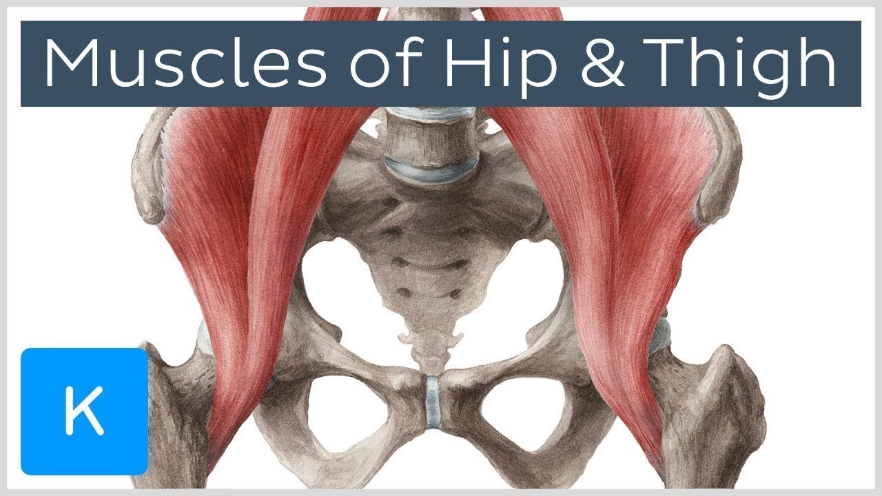
Muscles of the Hip and Thigh - Human Anatomy | Kenhub - ViDoe from i.ytimg.com The second largest bone in physique is the tibia, additionally known as the shinbone. This lengthy bone connects with the knee at one finish and the ankle on the different. A leg bone is a bone found in the leg. Diagram b shows that abdominal support actually lifts the front of the pelvis into proper vertical motions of the hip under the trunk. Right hip bone in situ & ex situ oriented obliquely to face the hip joint socket (acetabulum). The knee joint is the largest joint in the body and is primarily a hinge joint, although some sliding and rotation occur. Download this free vector about diagram showing the hip bone treatment, and discover more than 15 million professional graphic resources on freepik. These same nerves innervate the knee, which explains why pain can be referred to the knee from the hip and vice versa.
Each leg is made up of four bones.
Leg bones diagram femur manual e books. The hip joint gives the leg an incredible range of motion while still providing support to the body's weight. The head of your femur fits into your hip socket and the bottom end connects to your knee. The femur is the upper leg bone or thigh. It joins the lower limb to the pelvic girdle. Normally, a smooth cushion of shiny white hyaline (or articular) gluteus medius and minimus are the main abductors of the hip —that is, they move the leg away from the midline of the body (using the spine as a midline. Basic bone diagram enthusiast wiring diagrams. The knee is a strong but flexible hinge joint that uses muscles and. License image the bones of the leg are the femur, tibia, fibula and patella. In humans the neck of the femur connects the shaft and head at a 125 degree angle. Learn about hip and leg bones with free interactive flashcards. Bones of leg and foot. The foot bones shown in this diagram are the talus, navicular, cuneiform, cuboid, metatarsals and calcaneus.
Each leg is made up of four bones. The foot bones shown in this diagram are the talus, navicular, cuneiform, cuboid, metatarsals and calcaneus. Hip anatomy, function and common problems. Front view of the hip joint bones. Label bone diagram leg bone hip bone anatomy
12 Best Images of Skull Anatomy And Physiology Worksheets ... from www.worksheeto.com Fibula and tibia, ankle and foot. Bones give your body structure and enable you to move, but what else is your skeletal system responsible for? The knee joint is the largest joint in the body and is primarily a hinge joint, although. In humans the neck of the femur connects the shaft and head at a 125 degree angle. Each leg is made up of four bones. Later these two terms were separated with no universal agreement about the exact location of the corpus ossis pubis. On top of that layer of muscle is the iliotibial band, which starts at the brim of your pelvis outside the hip joint and runs down your leg. Labeled skeleton diagram best of pelvic bones simple bone diagram.
On top of that layer of muscle is the iliotibial band, which starts at the brim of your pelvis outside the hip joint and runs down your leg.
Feet human anatomy bones tendons ligaments and more. Download this free vector about diagram showing the hip bone treatment, and discover more than 15 million professional graphic resources on freepik. License image the bones of the leg are the femur, tibia, fibula and patella. Click and start learning now! The knee is a strong but flexible hinge joint that uses muscles and. These same nerves innervate the knee, which explains why pain can be referred to the knee from the hip and vice versa. Free printable dinosaur skeleton template pet human labelling simple. Learn how to to left from and right and the meaning behind the names of the. Find the perfect bone diagram stock illustrations from getty images. The hip joint gives the leg an incredible range of motion while still providing support to the body's weight. Quizzes on human skeletal system anatomy, bone anatomy, and bone. A diagram of the human skeleton. Front view of the hip joint bones.
Leg bones diagram femur manual e books. The head of your femur fits into your hip socket and the bottom end connects to your knee. Femur, upper bone of the leg or hind leg. The bone surfaces of the femoral head and acetabulum have a smooth durable layer of articular cartilage that cushions the ends of the bones and allows for smooth movement. When you stand or walk, all the weight of your upper body rests on them.
Diagram of the bones of the right leg and hip - Diagram of ... from www.mediastorehouse.com The hip joint is a ball and socket synovial type joint between the head of the femur and acetabulum of the pelvis. The pelvis and the femur (the thighbone). It joins the lower limb to the pelvic girdle. When you stand or walk, all the weight of your upper body rests on them. Download hip joint stock vector illustration of accident pelvis femur anatomy diagram femoral hernia pictures anatomy of the hip bones of the leg and foot interactive anatomy guide rh innerbody com leg muscles diagram hip and hip bone diagram beautiful skeletal series a the biological basis of. Each leg is made up of four bones. The knee joint is the largest joint in the body and is primarily a hinge joint, although some sliding and rotation occur. At the distal end of the femur, two rounded condyles meet the tibia and fibula bones of the lower leg to form the knee joint.
The knee is a strong but flexible hinge joint that uses muscles and.
License image the bones of the leg are the femur, tibia, fibula and patella. Feet human anatomy bones tendons ligaments and more. At the distal end of the femur, two rounded condyles meet the tibia and fibula bones of the lower leg to form the knee joint. Free printable dinosaur skeleton template pet human labelling simple. Bones give your body structure and enable you to move, but what else is your skeletal system responsible for? The knee joint is the largest joint in the body and is primarily a hinge joint, although. Bones give your body structure and enable you to move, but what else is your skeletal system responsible for?. Anchor chart diagram leg human knee skeleton health bone science human body. Hip anatomy, function and common problems. Find the perfect bone diagram stock illustrations from getty images. The foot bones shown in this diagram are the talus, navicular, cuneiform, cuboid, metatarsals and calcaneus. Download this free vector about diagram showing the hip bone treatment, and discover more than 15 million professional graphic resources on freepik. Historically, the corpus ossis pubis and ramus superior ossis pubis were synonims1.
At the distal end of the femur, two rounded condyles meet the tibia and fibula bones of the lower leg to form the knee joint leg bone diagram. Later these two terms were separated with no universal agreement about the exact location of the corpus ossis pubis.


0 Komentar